Light-Ultrasound Imaging Detects Thyroid Cancer Without Biopsy

Domestic researchers have developed a new imaging system that combines light and ultrasound to more accurately distinguish thyroid cancer (thyroid cancer) without the need for a biopsy. When a nodule is found in the thyroid, most patients must undergo a biopsy using a needle, but this new technology is expected to significantly reduce that burden.
Professor Kim Chulhong from POSTECH’s Department of Electrical and Electronic Engineering, IT Convergence Engineering, Mechanical Engineering, and the Graduate School of Convergence, along with Professor Lim Dong-jun, Professor Lee Jae-kyung from Catholic University of Korea Seoul St. Mary’s Hospital, and Professor Park Byul-ri from Sungkyunkwan University, jointly published these findings in the international journal *Science Advances* on August 27 of last year.
Typically, thyroid cancer diagnosis begins with an ultrasound examination. If a suspicious nodule is detected, a biopsy is performed using a needle to collect tissue samples. However, ultrasound alone has low accuracy in distinguishing between benign and malignant nodules, leading to unnecessary biopsies for non-cancerous nodules. This places physical and psychological burdens on patients and raises concerns about diagnostic accuracy among medical staff.
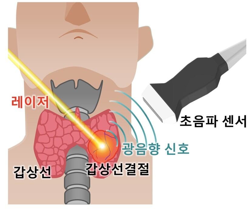
The research team, supported by the POSTECH-Catholic University of Korea Institute for Biomedical Science and Technology (POGA Research Institute), developed a “photoacoustic imaging (PAI)” technology. Malignant nodules are metabolically active, resulting in lower oxygen saturation. By measuring blood oxygen saturation through subtle ultrasound signals emitted by red blood cells when exposed to a laser (light), the system determines whether a nodule is benign or malignant. However, this method alone had limitations in distinguishing various types of thyroid cancer.
The team utilized data from 106 patients: 45 with papillary thyroid cancer, 32 with follicular tumors, and 29 with benign nodules. They extracted parameters such as oxygen saturation, asymmetry of distribution, and spectral slope from photoacoustic images and analyzed them using machine learning (AI) to develop a new diagnostic system called the “ATA-Photoacoustic (ATAP) score.”
The results showed a sensitivity of 97% in detecting malignant nodules, while specificity—the ability to exclude benign nodules from unnecessary testing—increased to 38%, more than double the 17% achieved by conventional ultrasound. This reduction in unnecessary tests could alleviate patient burden and lead to cost savings in medical expenses.
Professor Kim Chulhong stated, “This research is significant as it combines photoacoustic and ultrasound imaging to distinguish malignancies, including follicular tumors, which were previously difficult to diagnose.” Professor Park Byul-ri added, “Through follow-up research, we aim to enhance the technology’s completeness and conduct large-scale clinical validation to develop it into a medical device for practical use in healthcare settings.”
Reference
Science Advances (2025), DOI: https://doi.org/10.1126/sciadv.ady6173


Comments
Post a Comment